Phytoplasmas, the Fast Spreading Vector Borne Pathogens of Flower Crops: Indian Scenario
DOI:
https://doi.org/10.55446/IJE.2022.453Keywords:
Floriculture, disease, diagnosis, genomics, managementAbstract
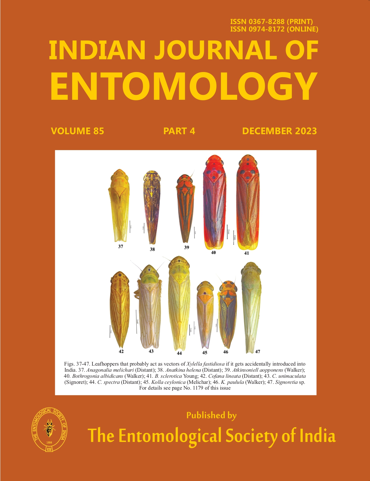
Floriculture trade in India is one of the most lucrative fields, earning valuable forex to the national exchequer. Exchange of planting material via different modes plays a major role in spreading of the pathogen. Floriculture being an international industry, the transmission of pests or pathogens through live plants, seeds, cuttings and flowers etc., is very high. In recent years, the occurrence of phytoplasmal diseases in flower crops is increasing due to extensive exchange amongst many groups. The prominent symptoms of phytoplasma infection are the flowers turning into leafy structures called phyllody and the formation of sterile green flowers called virescence. In floricultural crops, these two symptoms are of major disadvantages as the flowers are the main economic product. The phyllody symptoms are most often mistaken for novel plant type and are multiplied unwittingly. Phytoplasmas are switch between plant and animal kingdoms. Phytoplasma spread in the field through leaf hoppers by inhabiting in their gut, haemolymph, salivary glands etc. Among the ornamental plants and flower crops, various groups of phytoplasma have been reported from India. More than 40 per cent of the infections are reported from the members of the Asteraceae family with 16SrI-B group, ‘Candidatus Phytoplasma asteris’.
Downloads
Metrics
Downloads
Published
How to Cite
Issue
Section


References
Ajayakumar P V, Samad A, Shasany A K, Gupta A K, Alam M,Rastogi S. 2007. First record of a ‘Candidatus phytoplasma’ associated with little leaf disease of Portulaca grandiflora. Australian Plant Disease Notes 2: 67-69.
Alma A, Bosco D, Danielli A, Bertaccini A, Vibio M. 1997. Identification of phytoplasmas in eggs, nymphs and adults of Scaphoideustitanus Ball reared on healthy plants. Insect Molecular Biology 6:115-121.
Arocha-Rosete Y, Singh A, Pandey M, Tripathi A N, Chandra B, Shukla S K, Singh Y, Kumar A, Srivastava R K, Zaidi N W, Arif M, Narwal S, Tewari A K, Gupta M K, Nath P D, Rabindran R, Khirbat S K, Byadgi A S, Singh G, Boa E. 2008. New plant hosts for group 16SrII, ‘Candidatus phytoplasma aurantifolia’ in India. Plant Pathology 17: 36.
Bai X D, Zhang J H, Ewing A, Miller S A, Radek A J, Shevchenko D V. 2006.Living with genome instability: the adaptation of phytoplasmas to diverse environments of their insect and plant hosts. Journal of Bacteriology 188 (10): 3682-3696.
Beanland L, Hoy C W, Miller S A, Nault L R. 2000. Influence of aster yellows phytoplasma on the fitness of the aster leafhopper (Homoptera: Cicadellidae). Annals of Entomological Society of America 93:271-276.
Bertaccini A, Lee IM. 2018. Phytoplasmas: an update in: Phytoplasmas: Plant pathogenic bacteria-I. Characterization and epidemiology of phytoplasma associated diseases.Singapore: Springer pp. 1-29.
Chaturvedi Y, Tiwari A K, Upadhyaya P P, Prabhuji S K, Rao G P. 2009a. Association of ‘Candidatus phytoplasma asteris’ with little leaf and phyllody disease of Catharanthus roseus in Eastern Uttar Pradesh, India. Medicinal Plants 1: 103-108.
Chaturvedi Y, Singh M, Rao G P, Snehi S K, Raj S K. 2009b. First report of association of ‘Candidatus Phytoplasma asteris’ (16SrI group) with little leaf disease of rose (Rosa alba) in India. Plant Pathology 58: 788.
Chaturvedi Y, Singh M, Snehi SK, Raj SK, Rao GP. 2010. First report of ‘Candidatus Phytoplasma asteris’ (16SrI group) associated with yellows and little leaf diseases of Hibiscus rosa-sinensis in India. Plant Pathology 59: 796.
Cho S, Lin C, Kuo C. 2019. Genomic characterization of the periwinkle leaf yellowing (PLY) phytoplasmas in Taiwan. Frontiers in Microbiology 10: 2194.
Dickinson M. 2015. Loop-mediated isothermal amplification (LAMP) for detection of phytoplasmas in the field. Methods in Molecular Biology 1302:99-111.
Doi Y, Terenaka M, Yora K, Asuyama H. 1967. Mycoplasma or PLT group-like microorganisms found in the phloem elements of plants infected with mulberry dwarf, potato witches’ broom, aster yellows, or paulownias witches’ broom. Annals Phytopathological Society Japan 33: 259-266.
Gopala G P. 2018. Molecular characterization of phytoplasma associated with four important ornamental plant species in India and identification of natural potential spread sources. 3 Biotech 8:116.
Gundersen D E, Lee IM, Schaff D A, Harrison N A, Chang C J, Davis R E, Kinsbury D T. 1996. Genomic diversity among phytoplasma strains in 16S rRNA Group I (Aster Yellows and related phytoplasmas) and III (X-Disease and related phytoplasmas). International Journal of Systematic Bacteriology 46: 64-75.
IRPCM. 2004. Candidatus phytoplasma, a taxon for the wall-less, non-helical prokaryotes that colonizeplant phloem and insects. International Journal of Systematic and Evolutionary Microbiology 54: 1243-1255.
Kawakita H, Saiki T, Wei W, Mitsuhashi W, Watanabe K. 2000. Identification of mulberry dwarf phytoplasmas in the genital organs and eggs of leafhopper Hishimonoides sellatiformis. Phytopathology 90: 909-914.
Khasa E, Taloh A, Gopala PT, Prabha K, Rao GP. 2016. Molecular characterization of phytoplasmas of clover proliferation group associated with three ornamental plant species in India. 3 Biotech 6 (2): 1-7.
Kirdat K, Tiwarekar B, Thorat V, Narawade N, Dhotre D, Sathe S, Shouche Y, Yadav A. 2020. Draft genome sequences of two phytoplasma strains associated with sugarcane grassy shoot (SCGS) and bermuda grass white leaf (BGWL) diseases. Molecular Plant-Microbe Interactions, 33(5): 715-717.
Kumar S, Jadon V, Towari A K, Rao G P. 2015. Exitianus indicus (Distant): a putative vector for ‘Candidatus Phytoplasma cynodontis’ in India. 5 (1-Supplement): S51 - S52.
Linck H, Lankes C, Kruger E, Reineke A. 2019. Elimination of Phytoplasmas in Rubus Mother Plants by Tissue Culture Coupled with Heat Therapy. Plant Disease Notes 103(6): 1252-1255.
Madhupriya, Rao G P. Khurana S M P. 2015. Rice yellow dwarf phytoplasma (16Sr XI-B subgroup) infecting Jasminum sambac in India. Phytoparasitica 43: 77-80.
Manish Kumar, Madhupriya, Rao, G. P. 2017. Molecular characterization, vector identification and sources of phytoplasmas associated with brinjal little leaf disease in India. 3 Biotech 7:7.
Nayar R. 1977. Sandal spike disease and control of spike [India]. Journal of the Indian Academy of Wood Science (India) 8(1): 41-49.
Oshima K, Kensaku M, Namba S.2013. Genomic and evolutionary aspects of phytoplasmas. Frontiers in Microbiology 4: 230.
Panda P, Debnath P, Mall S, Nigam A, Rao G P. 2021. Multilocus genes based characterization of phytoplasma strains associated with Mexican and french marigold species in India. European Journal of Plant Pathology 161(2): 313-30.
Panda P, Nigam A, Rao G P. 2021. Multilocus gene analysis reveals the presence of two phytoplasma groups in Impatiens balsamina showing flat stem and phyllody. 3 Biotech 11:122.
Pathak D M, Parakhia A M, Akbari L F. 2012. Symptomatology and transmission of sesame phyllody disease caused by phytoplasma. Journal of Mycology and Plant Pathology 42:479-484.
Prabha K, Nitika Gupta, Girish K S, Prasad K V. 2017. Phytoplasma in nurseries: An imminent threat to floriculture. Nursery Today. pp. 30-36.
Prabha K, Gupta N, Girish K S, Kadam G B, Saha T N, Prasad K V. 2019. Molecular characterization of phytoplasma associated with members of daisy family based on 16SrRNA. In XIth National Symposium of Indian Association of Mycoplasmologists -International Conference on Human, Animal and Plant Mycoplasmas, from 3rd to 4th December 2019. Abstract. pp. 97.
Purcell AH. 1988. Increased survival of Dalbulus maidis, a specialist on maize, on non-host plants infected with mollicute plant pathogens. Entomologia Experimentaliset Applicata 46: 187-196.
Raj SK, Khan MS, Kumar S. 2007. Molecular identification of ‘Candidatus phytoplasma asteris’ associated with little leaf disease of Chrysanthemum morifolium. Australasian Plant Disease Notes 2: 21-22.
Raj S K, Snehi S K, Kumar S, Banerji B K, Dwivedi A K, Roy R K, Goel A K. 2009. First report of ‘Candidatus Phytoplasma asteris’ (16SrI group) associated with colour-breaking and malformation of floral spikes of gladiolus in India. Plant Pathology 58: 1170.
Raj S K, Snehi S K, Khan M S, Singh M, Chaturvedi Y, Tiwari A K, Rao G P. 2011. Molecular detection and identification of phytoplasma associated with little leaf and witches’ broom disease of marigold in India. Phytopathogenic Mollicutes 1(1): 41-46.
Rao G P, Madhupriya, Thorat V, Manimekalai R, Tiwari A K, Yadav A. 2017. A century progress of research on phytoplasma diseases in India. PhytopathogenicMollicutes7 (1): 1-38.
Rao G P, Madhupriya, A K Tiwari S, Kumar, V K Baranwal. 2014. Identification of sugarcane grassy shoot-associated phytoplasma and one of its putative vectors in India. Phytoparasitica 42: 349-354.
Rihne T, Kumar M, Shreenath Y S, Pant R P, Taloh A, Swaroop K, Rao G P. 2019. Mixed infection of virus and phytoplasma in gladiolus varieties in India. Phytopathogy Mollicutes 9(1): 149-150.
Rihne T, Mitra S, Bahadur A, Panda P, Banyal N, Rao G P. 2020. Mixed infection of phytoplasma and begomovirus associated with leaf curling and witches’ broom disease of Zinnia elegans in India. Indian Phytopathology 73(3): 527-532.
Sajad un Nabi, Madhupriya, Dubey D K, Rao G P, Baranwal V K, Pratibha Sharma. 2015. Molecular characterization of ‘Candidatus Phytoplasma asteris’ subgroup I-B associated with sesame phyllody disease and identification of its natural vector and weed reservoir in India.
Shukla K, Madhupriya, Dubey D, Upadhyaya PP, Rao GP. 2014. Association of pigeon pea witches’ broom phytoplasma. Infecting Carpobrotus edulis (L.) N.E. Br in India. Phytopathogenic Mollicutes 4: 27-32.
Singh S K, Amminudin, Srivastava P, Singh BR, Khan J A. 2007. Production of phytoplasma-free plants from yellow leaf diseased CatharanthusroseusL. (G.) Don. Journal of Plant Diseases and Protection 114: 2-5.
Singh M, Chatruvedi Y, Tiwari A K, Rao GP, Snehi S K, Raj S K, Khan M S. 2011. Diversity among phytoplasmas infecting ornamental plants grown in India. Bulletin of Insectology 64(Supplement): S69-S70.
Srivastava S, Singh V, Gupta P S, Sinha O K, Baitha A. 2006, Nested PCR assay for detection of sugarcane grassy shoot phytoplasma in the leafhopper vector Deltocephalus vulgaris: A first report. Plant Pathology 22: 25-32.
Sumashri K S, Yadav V, Kirdat K, Vipool T, Bhavesh T, Yadav A, Gottravalli R. 2020. Molecular detection and identification of a strain related to Candidatus phytoplasma australasia’ in marigold in India. Phytopathogenic Mollicutes 10(1): 82-88.
Taloh A, Raju D V S, Namita Banyal, Gunjeet Kumar, Priyam Panda, Manimekalai R, Marcone C, Rao G P. 2020. Genetic diversity of phytoplasma strains infecting chrysanthemum varieties in India and their possible natural reservoirs. 3Biotech 10:411.
Tiwari A K, Tripathi S, Lal M, Sharma M L, Chiembostat P. 2011. Elimination of sugarcane phytoplasma through apical meristem culture. Archives of Phytopathology and Plant Protection 44 (20):1942-1948.
Tiwari A K, Madhupriya, Srivastava V K, Pandey K P, Sharma B L, Rao G P., 2016a. Detection of sugarcane grassy shoot phytoplasma (16SrXI-B subgroup) in Pyrillaperpusilla Walker in Uttar Pradesh, India. Phytopathogenic Mollicutes 6 (1): 56 - 59.
Tiwari A K, Shailender Kumar, Smriti Mall, Vikas Jadon, Rao G P. 2016b. New efficient natural leafhopper vectors of Sugarcane grassy shoot phytoplasma in India. Sugar Tech 19(2):191-197.
Weintraub P G, Pivonia S, Rosner A, Gera A. 2004. A new disease in Limonium latifolium Hybrids. II. Investigating insect vectors. Hort Science 39(5): 1060-1061.